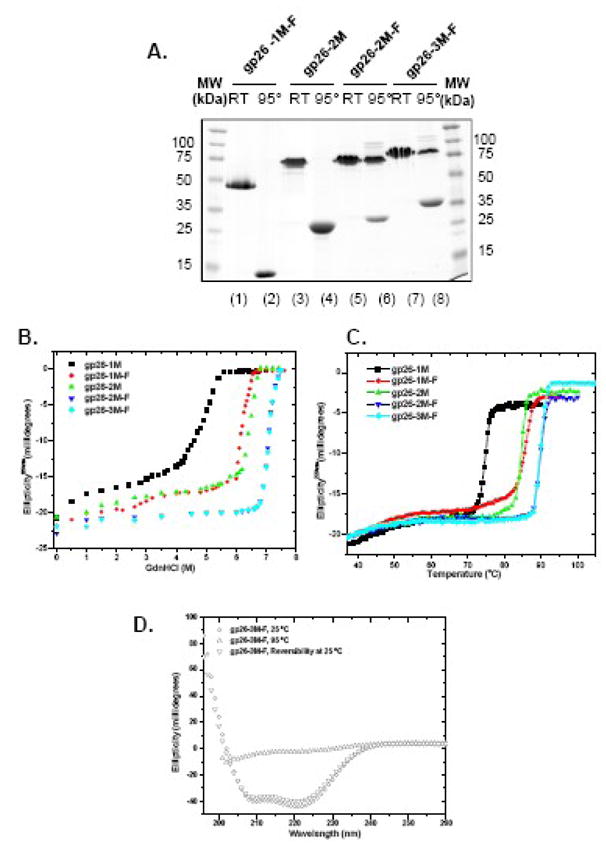
Figure 7. In frame insertion of MiCRUs in gp26-Foldon chimeric fibers.

(A) SDS-PAGE analysis of gp26-MiCRU foldon fibers. “95°” and “RT” denote samples that were either boiled for 5 min or incubated 5 min at 22°C prior to be separated on gel. Stability of gp26-F-fibers was also measured independently against GdnHCl (B) and thermal (C) denaturation. (D) Reversibility of gp26-3M-F fiber was assessed by circular dichroism. The CD spectrum of fiber was measured between 197–260 nm in 20 mM sodium phosphate buffer pH 8 and 170 mM sodium chloride. Circular dichroism spectra was measured at 25°C, 95°C and after 30 minutes of cooling from 95 to 25°C (lower triangle). gp26-3M-F fiber refolded immediately after cooling. The protein concentration used in these experiments was 6μM.
