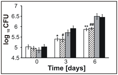
Figure 5. The viability of lysX in macrophages.
THP-1-derived macrophages were infected with Mtb strains; at the indicated times following infection, the macrophages were lysed, and the Mtb viability was determined. The white bars represent Rv-80lys; the dashed bars represent Rv-82med; the grey bars represent Rv-81ami; and the black bars represent Rv-03. Data are mean±standard error from three independent experiments, and the Mann-Whitney Rank Sum Test was used for data analyses.

