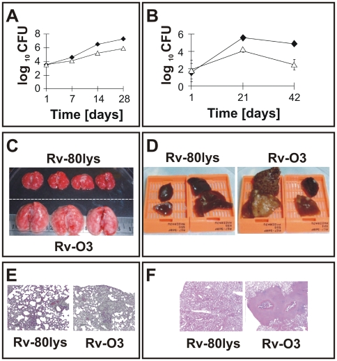
Figure 8. Growth of lysX and WT strains in vivo.
Panel A: C57BL/6 female mouse lungs were infected with lysX (triangles) and WT (dark diamonds) strains via an aerosol route. The mean CFU counts were obtained from lung homogenates of at least three mice per group and plated on Middlebrook 7H10 agar plates. All plates were incubated at 37°C for at least three weeks before colonies were counted. Panel B: Guinea pigs (Hartley strain) were infected with lysX (open symbols) or WT (dark symbols) strains via an aerosol route. Five guinea pigs were sacrificed at days 1, 21 and 42 days after infection, and the survival of Mtb strains in the entire lung homogenate was determined by plating on agar medium as described above. Panels C, D: Gross pathology of the lungs infected with the Rv-80lys and Rv-03 Mtb strains. Lungs from mouse -C- and guinea pig -D- were excised, stored in 10% formalin, embedded, and stained with hematoxylin and eosin for histopathological analysis. Note the presence of tubercles on the surface of the lungs for Rv-03 compared to Rv-80lys. Panels E, F: Histopathology of mouse -E- and guinea pig -F- lungs infected with the Rv-80lys and Rv-03 Mtb strains. Hematoxylin-eosin staining confirmed that the lungs infected with Rv-03, but not those infected with Rv-80lys, had extensive inflammation in both species and showed caseating granulomas in guinea pigs.

