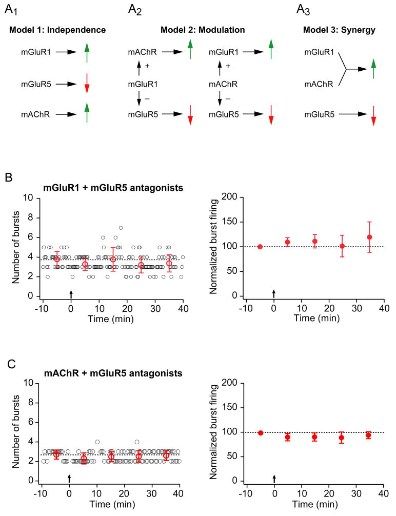
Figure 5. Synergistic activation of mGluR1 and mAChRs is required for enhancement of burst firing while activation of mGluR5 mediates suppression of burst firing.
A. Three models for the effects of metabotropic receptor activation on burst firing. A1. The first model that accounts for the effects of mGluR1, mGluR5, and mAChR on the induction of burst plasticity is based on the results of pharmacological experiments with metabotropic receptor antagonists (see Fig. 4). In Model 1, each receptor has an independent effect on burst firing: mGluR1 is necessary for an increase (because when blocked, a suppression of burst firing is observed), mGluR5 is necessary for a decrease (because when blocked, an enhancement of burst firing is observed), and mAChRs are necessary for an increase (because when blocked, a suppression of burst firing is observed). A prediction of Model 1 is that when both mGluR1 and mGluR5 are blocked, an increase in burst firing would be observed, mediated by mAChR activation alone. However, under these conditions (see panel B below) cells displayed no change in burst firing, demonstrating that this model does not account for the experimental results. A2. Model 2 proposes that mAChR or mGluR1 has a modulatory effect on the other two receptors, enhancing an mGluR1 (or mAChR) -mediated increase or inhibiting an mGluR5-mediated decrease in burst firing (or both). A prediction of Model 2 is that when mGluR5 is blocked with either mGluR1 or mAChR, an increase in burst firing would be observed due to the action of mAChR or mGluR1 alone. However, no change in burst firing was observed in these conditions (see panels B and C), demonstrating that this model can not account for the observed plasticity. A3. A third possiblitiy is that both mGluR1 and mAChR must be activated together to induce an enhancement of burst firing (and mGluR5 activation alone leads to a suppression). Model 3 predicts that when mGluR5 is blocked in combination with either mGluR1 or mAChR, no increase in burst firing should be observed. These predictions are consistent with the observed results. B. Representative (left) and group (right; n=8) data from experiments in which TBS was given in the presence of a specific mGluR1 antagonist (LY367385, 25 μM) and a specific mGluR5 antagonist (MPEP, 10 μM). For the representative-experiment graph, small open circles (black) indicate the number of burst firing responses evoked by a train of 10 EPSC-like somatic current injections. The train was delivered every 20 seconds. Large open circles (red) represent the average number of burst firing responses per train for each 10-minute period. Error bars are ± standard deviation. For the group-data graph, filled symbols represent the average number of burst firing responses per train for each 10-minute period. Error bars are ± s.e.m. For both graphs, dotted lines indicate the average number of burst firing responses per train for the 10-minute baseline period. Arrows indicate when TBS (induction) was given. C. Representative (left) and group (right; n=6) data from experiments in which TBS was given in the presence of an mAChR antagonist (atropine, 10 μM) and a specific mGluR5 antagonist (MPEP, 10 μM). Layout is as described in B.

