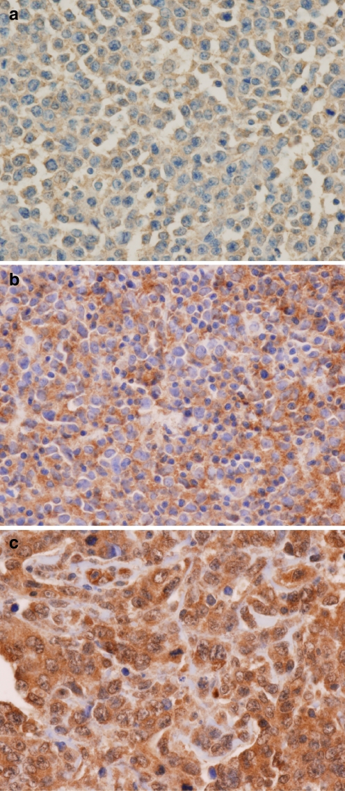
Fig. 1.
Representative photomicroghs of c-REL expression in DLBCL (original magnification, ×400; immunohistochemical c-REL stain). a, b Negative c-REL expression cases: Negative nuclear expression with weak (a) and moderate (b) cytoplasmic expression, respectively. c Case scored as positive c-REL showing both nuclear and cytoplasmic staining; note small lymphocytes without nuclear or cytoplasmic staining

