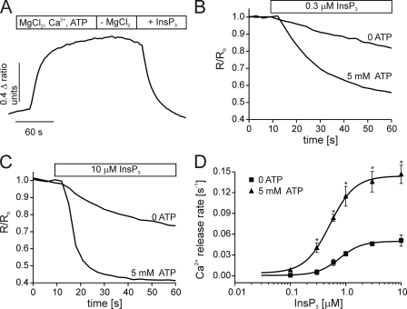
FIGURE 1.
ATP enhances IICR from DT40-3KO cells stably expressing rat InsP3R1. ER luminal Ca2+ measurements were obtained from cells stably expressing rat S2+ InsP3R1. Cells were loaded with furaptra and permeabilized with β-escin as described under “Experimental Procedures.” A shows the experimental paradigm. ER stores are loaded with Ca2+ by superfusion with a solution containing Mg2+, Ca2+, and ATP. A solution lacking Mg2+ is then applied to prevent SERCA activity. InsP3 initiates the release of Ca2+ from the ER. B and C show representative traces from cells treated with low (0.1 μm, in B) or high (10 μm, in C) InsP3 in the presence or absence of 5 mm ATP. D shows the concentration-response relationship for InsP3 in the absence (squares) or presence (triangles) of 5 mm ATP. Each point is the mean ± S.E. of at least four separate experiments. The solid lines are the fits of the data. Ca2+ release rates were calculated by fitting the average time course from the first 30 s of InsP3 application from 20–50 cells to a single exponential. (*, p ≤ .05; Student's unpaired t test).

