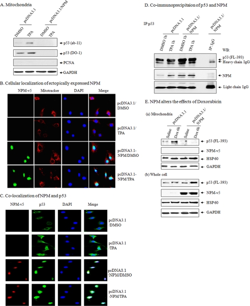
FIGURE 2.
NPM suppresses mitochondrial p53 levels. A, suppression of mitochondrial p53 following NPM overexpression in isolated purified mitochondria following DMSO or TPA treatment for 1 h in JB6 cells. B, localization of NPM following NPM overexpression using antibody specific for exogenously expressed NPM (anti-NPM-V5 antibody) staining and confocal microscopy. Green, NPM; red, MitoTracker; blue, nuclear marker 4′,6-diamidino-2-phenylindole (DAPI). C, co-localization of NPM with p53 using NPM and p53 antibody staining and confocal microscopy. Green, p53; red, NPM (anti-NPM-V5 antibody); blue, 4′,6-diamidino-2-phenylindole. D, interaction of p53 and NPM in JB6 cells. E, overexpression of NPM suppresses mitochondrial p53 in isolated purified mitochondria with doxorubicin treatment in JB6 cell mitochondria (a) and total cell lysates (b). GAPDH, glyceraldehyde-3-phosphate dehydrogenase. IP, immunoprecipitation; WB, Western blot.

