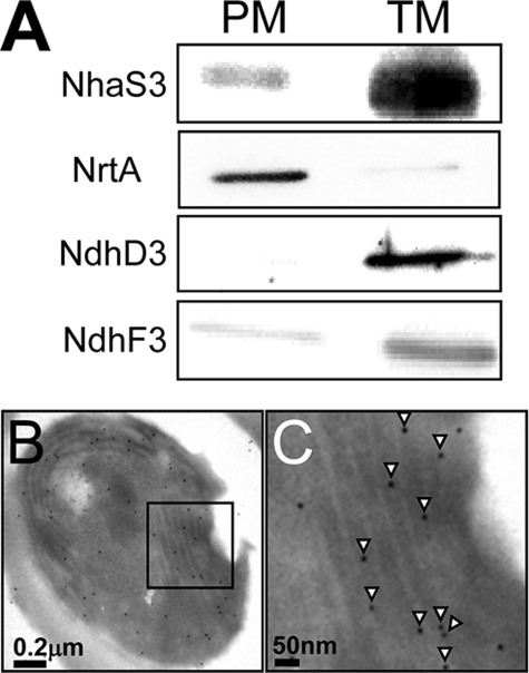
FIGURE 2.
Membrane localization of NhaS3. A, membrane fractions were prepared by aqueous polymer two-phase partitioning and sucrose density gradient and separated by SDS-PAGE. NhaS3, NrtA, NdhD3, and NdhF3 proteins were detected on Western blots using the corresponding antibodies. PM, plasma membrane; TM, thylakoid membrane. B and C, cross-section of a Synechocystis cell immunolabeled using an anti-NhaS3 antibody. C is an enlarged section of B. NhaS3 protein, indicated by the presence of gold particles (arrowheads in C), was detected in the thylakoid membrane.

