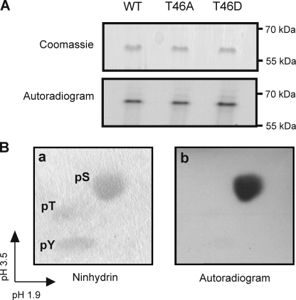
FIGURE 2.
In vitro phosphorylation of Kv1.2 serine residue(s) by PKA. A, Kv1.2-WT-myc, -T46A-myc, and -T46D-myc immunoprecipitates treated with cPKA in the presence of [γ-32P]ATP and visualized by Coomassie Blue staining and autoradiography to detect 32P incorporation. Data are representative of six experiments. B, phosphoamino acid analysis of phosphorylated Kv1.2-WT-myc protein by two-dimensional electrophoresis. Unlabeled phosphoserine (pS), phosphothreonine (pT), and phosphotyrosine (pY) standards were detected by ninhydrin staining (a) and 32P-labeled residues by autoradiography (b). Data are representative of four experiments.

