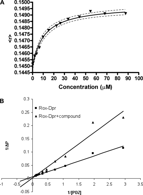
FIGURE 3.
Binding of compound 3289-8625 to the Dvl PDZ domain. A, binding was followed by monitoring the changing of fluorescence anisotropy of the TMR-labeled Dvl PDZ domain by binding compound 3289-8625. In the plot of the fluorescence anisotropy of the TMR-labeled Dvl PDZ domain with increasing concentrations of compound 3289-8625, the y axis is fluorescence anisotropy, and the x axis is the concentration of the compound. The Kd value was determined from the fitted curve. B, polarization change was monitored during titration of the PDZ domain into 50 nm ROX-Dpr peptide solution in the absence and presence of 6 μm compound 3289-8625, respectively; the reciprocal of the polarization change (1/ΔP) was plotted against the reciprocal of the PDZ protein concentration (1/[PDZ]). Data were analyzed by the program Prism (GraphPad Software Inc.).

