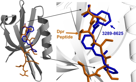
FIGURE 4.
The complex structures. The crystal structure of the Dvl PDZ domain-bound Dpr peptide is shown overlaid with the highest scoring predicted conformation (from FlexX) of compound 3289-8625 in the binding groove. The carbon atoms of the bound Dpr peptide are shown in brown, the carbon atoms of the bound compound 3289-8625 are shown in blue, and the carbon atoms of the PDZ domain are shown in gray.

