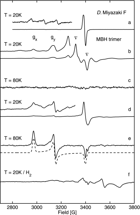
FIGURE 5.
EPR spectra of R. eutropha MBH attached to the membrane (traces b–f) and oxidized (as isolated) D. vulgaris Miyazaki F hydrogenase (trace a). The enhanced (trace a) shows signals from superimposed gx and gy components of Niu-A and Nir-B. Experimental conditions are as follows: 1 milliwatt microwave power; 1 mT modulation amplitude, 12.5 kHz modulation frequency. Trace b, EPR spectra of oxidized (as isolated or reoxidized) MBH from a preparation of the cytoplasmic membrane at T = 20 K (redox potential +290 mV) showing signals from Nir-B and split signal from [3Fe4S]+. Trace c, oxidized (as isolated) from cytoplasmic membrane preparations at T = 80 K. Trace d, MBH, partially reduced with 5 mm β-mercaptoethanol (+40 mV), recorded at T = 20 K (the complex split [3Fe4S] signal has disappeared, and a narrow signal arises at a redox potential of +40 mV), Enhanced trace, Nir-B. Trace e, MBH partially reduced with 5 mm β-mercaptoethanol (+40 mV), recorded at T = 80 K; the dashed line represents the simulated spectrum of Nir-B. Trace f, H2-reduced samples from cytoplasmic membrane preparation (redox potential −390 mV). Experimental conditions for spectra (traces b–f) are as follows: 10 milliwatt microwave power; microwave frequency 9.56 GHz; 1 mT modulation amplitude, 12.5 kHz modulation frequency.

