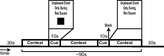
Figure 1.
Example of CBF image acquisition trial. Each trial followed an instructed threat procedure in which one of three conditions (N, P, or U) was presented for ∼1.5 min. Each condition was preceded and followed by 30 s of filler that was not included in analyses. In the N condition, no unpleasant events (shocks) were delivered. In the P condition (shown), unpleasant events were administered only in the presence of a threat cue. In the U condition, unpleasant events were delivered at any time. During each trial, two 10-s-duration cues were presented. Cues consisted of different colored shapes (color omitted here). The cues signaled the possibility of receiving an aversive stimulus in the P condition but had no signal value in the other conditions. Throughout each trial, a computer monitor apprised participants of the current condition by displaying one of the following messages: “No Unpleasant Event” (neutral), “Unpleasant Event Only During Red Square” (predictable), or “Unpleasant Event at Any Time” (unpredictable). This information, in the absence of a cue, constituted the context for each condition. One shock was administered during each trial involving a P or U condition. The shocks were delivered simultaneously with the offset of a cue in the P condition and in the absence of the cue in the U condition. Two CBF images were acquired in each condition: one when the cue was displayed along with the instructions and another when the cue was absent (i.e., during the context only). Thus, one CBF scan was acquired per trial.

