
Figure 5. Ubiquitylation of drICE Suppresses Its Catalytic Potential.
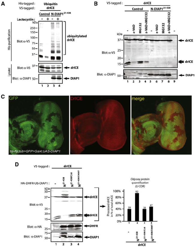
(A) Inhibition of the proteasome does not alter the ubiquitylation pattern of drICE. 293T cells were cotransfected with the indicated constructs and treated with Lactacystin or DMSO (control). The presence of ubiquitylated drICE was assayed as in Figure 2E.
(B) Proteasome inhibition failed to restore the appearance of the small subunit of drICE. S2 cells were cotransfected with the indicated constructs. Following a 2 hr drICE induction, cells were treated for a further 4 hr with MG132 or DMSO.
(C) Clonal expression of DIAP1 in the developing eye does not reduce the levels of drICE. Eye discs overexpressing DIAP1 in clones (marked by the absence of GFP, first and last panel) were stained using an α-drICE antibody (red, middle panel).
(D) S2 cells were cotransfected with the indicated constructs. Immunoblot analysis (left panel) revealed that zymogenic ALG-drICE accumulated at the expense of processed drICE (p10) in the presence of DIAP1. LI-COR Odyssey quantification (right panel) of the drICE signal.