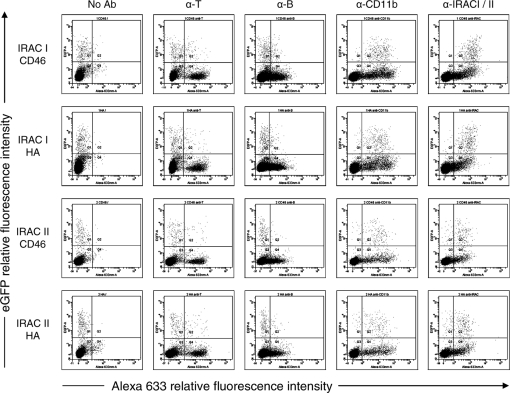
Figure 4.
Membrane anchored forms of IRAC proteins are detectable at the surface of PBMC infected ex vivo. Rabbit PBMC were collected as described in the methods and infected at a MOI of 1 PFU/cell with BoHV-4 V. test BAC G IRAC I CD46, V. test BAC G IRAC I HA, V. test BAC G IRAC II CD46 or V. test BAC G IRAC II HA strains. After 48 h, cells were treated as described in the methods for indirect immunofluorescent labeling with mAbs directed against rabbit CD11b, rabbit IgMs (α-B) or rabbit CD5 (α-T) and then analyzed by flow cytometry for eGFP and Alexa-633 fluorescences.

