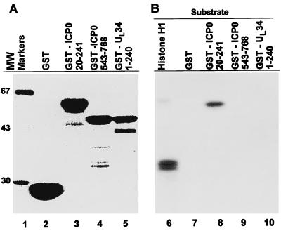
Figure 3.
(A) Coomassie blue staining of GST fusion proteins. GST alone, GST-ICP0 20–241, GST-ICP0 543–768, and UL34 1–240 were purified with the aid of glutathione-agarose beads, electrophoretically separated on 10% bisacrylamide gels, and stained with Coomassie blue. (B) Autoradiographic image of cdc2-mediated phosphorylation of histone H1 or indicated GST fusion proteins. Cdc2 was immunoprecipitated from uninfected HeLa cells with cdc2 antibody, and histone H1 or GST fusion proteins were tested as substrates for cdc2. Reactions were electrophoretically separated on 10% bisacrylamide gels, transferred to nitrocellulose membrane, and exposed to film.

