Figure 4.
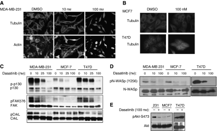
Dasatinib treatment disrupts cytoskeletal organisation and cellular invasion. (A, B) MDA-MB-231 (A), MCF7, and T47D (B) cells were grown on glass chamber slides and treated with dasatinib for 48 h before staining with antitubulin antibody and FITC-phalloidin. (C) MDA-MB-231, MCF7, and T47D cells were treated with increasing doses of dasatinib (DMSO, 10, 25, or 100 nM) for 2 h. Whole cell lysates were immunoblotted for phospho -FAK(Y576), -p130 (Y410) and -CrkL (Y207) . Membranes were then stripped and reprobed for total p130, FAK, or CrkL, respectively. (D) Cells were probed for phospho-N-WASP (then reprobed for total N-WASP) after treatment with DMSO, 10, or 100 nM dasatinib for 2 h. (E) DMSO- or dasatinib-treated cells were lysed and probed for phospho-Akt (S473). The membrane was then stripped and reprobed for total Akt as a control. All images are representative of at least three independent experiments.

