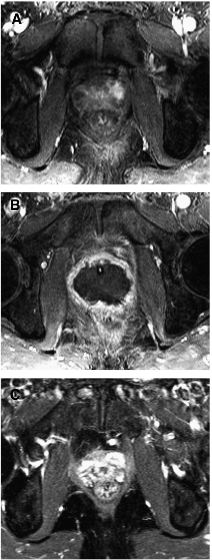
Figure 6.
Contrast enhanced MRI changes in a successful treatment for prostate cancer using HIFU. (A) 1.5 Tesla dynamic contrast enhanced MRI using gadolinium prior to HIFU treatment demonstrating localised disease with a lesion in the left antero-lateral side of the gland (circled). (B) 1.5 Tesla dynamic contrast enhanced MRI using gadolinium at 2 weeks demonstrating poor perfusion in the prostate after HIFU treatment. Urethral catheter seen in-situ. (C) 1.5 Tesla dynamic contrast enhanced MRI using gadolinium at 6 months no residual prostate tissue and fibrotic reaction.

