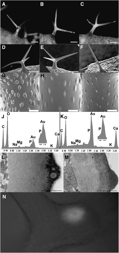
Figure 4.
Analysis of hdg2 Trichome Cell Wall Mutant.
(A–C) Stereomicrographs of trichomes from hdg2-2 mutant, wild-type, and rescued hdg2-2, respectively.
(D–F) SEM of wild-type, hdg2-2, and wild-type transformed with pEGAD MYB5:GFP–cHDG2 transgene showing co-suppressed hdg2-like mutant papillae phenotype, respectively.
(G–I) Higher magnification of trichomes shown in (D–F).
(J, K) Elemental analysis of wild-type and hdg2-2 trichome papillae.
(L, M) TEM of cross-section through branch of wild-type and hdg2-2 mutant trichomes.
(N) Localization of GFP–HDG2 fusion protein in wild-type plant transformed with the pEGAD MYB5:GFP–cHDG2 transgene.
Bars in (A–F) = 100 μm; (G–J) = 1 μm; (L, M) = 0.5 μm.

