
Fig. 4.
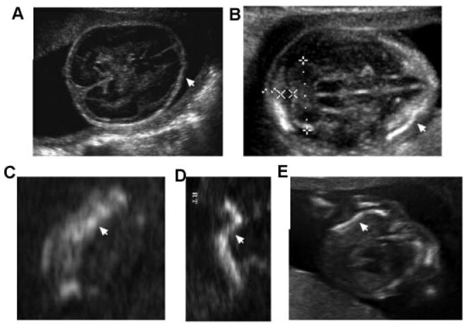
Sonographic images of osteogenesis imperfecta type 2 (OI type 2) showing abnormal findings. A: Absent mineralization of the calvarium (18 weeks gestation). B: Diminished mineralization of the calvarium (16 weeks gestation); arrow points to an area of calvarium mineralization. C: Femur (18 weeks gestation) showing bright areas (echodense) representing fracture and callus formation (arrow). D: Femur (16 weeks gestation) showing irregular bending (arrow). E: Saggital chest view in OI type 2 showing wavy or S-shaped ribs (arrow) and ribs that do not surround two-thirds of the chest circumference.