
Fig. 6.
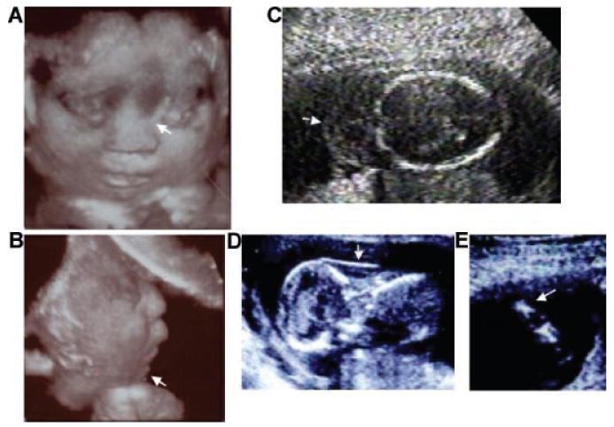
Sonographic images of achondrogenesis type 2. A,B: Three dimensional sonographic image of a fetus with achondrogenesis type 2 at 22 weeks gestation showing severe by flattened nasal bridge (arrow) and micrognathia (arrow). C: Cystic hygroma (arrow) at 20 weeks gestation. D: Increased nuchal fold (12 weeks gestation) (arrow) in the same fetus from (C). E: Femur (16 weeks gestation) showing severe micromelia and metaphseal spikes.