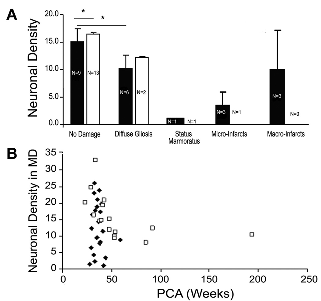
Figure 2.
A. Neuronal density in the four patterns of injury (Patterns 1–4) in PVL and control cases compared to the neuronal density in PVL and control cases without histopathologic changes (Pattern 0) in the mediodorsal nucleus. The neuronal density in Pattern 0 and Pattern 1 is presented as the mean adjusted for postconceptional age. There was significantly reduced neuronal density in the PVL cases compared to controls (p<0.05) in Pattern 0 in the mediodorsal nucleus, but not in the lateral posterior nucleus (data not shown). There was also a significant difference in neuronal density in the mediodorsal nucleus between Pattern 0 and Pattern 1 in PVL cases (p=0.001) but not controls (p=0.37). Neuronal density was markedly reduced in status marmoratus, micro-infarcts within infarct itself), and macro-infarcts. PVL, ■; Control, □. B. In PVL cases without status marmoratus or micro- or macro-infarcts, the neuronal density tended to decrease with increasing postconceptional age in both PVL and control cases in the mediodorsal nucleus, with a marginal difference in the mean density adjusted for postconceptional age in the PVL cases (11.9±1.6 neurons/mm2) compared to controls (16.7 neurons/mm2) (p=0.07). PVL, ◆; Control, □

