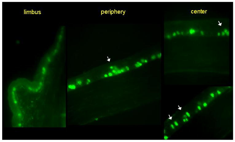
Figure 3.

Ki-67+ cells distribution among the corneal epithelium in mice subjected to desiccating stress (green). A significant number of Ki-67+ cells (34.2%) were detected in the suprabasal cell layers of the central and peripheral corneal epithelium (white arrows). No suprabasal cell was detected in the limbus of dry eye mice.
