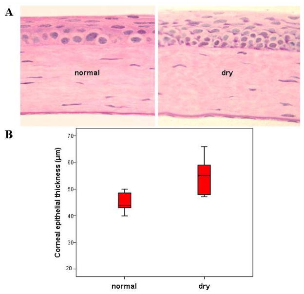
Figure 4.
Corneal epithelial thickness: comparison between normal and dry eye mice. A. Central corneal sections of 3 μm were stained with hematoxylin-eosin (H&E). Normal control cornea had a stratified epithelium of 4 to 6 cell layers and an avascular stroma with cells aligned in a lamellar manner. After 7 days of exposure to the CEC epithelial stratification was marked in the central regions. Besides marked stratification, the most striking finding was the increased cellularity and the general hyperplastic appearance of cells, evidenced by the cuboidal shape of postmitotic suprabasal cells (photographs were taken at the same magnification). B. Measurement of the central part of the corneal epithelium revealed that the epithelium was significantly thicker in eyes of dry eye mice as compared to the controls. Data show median ± interquartile; Mann-Whitney test.

