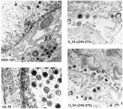
Figure 3.
Electron micrographs of cells infected with R5601(ΔUL34) or R5603 [UL34Δ(245–275)]. Rabbit skin cells exposed to 5 pfu of R5601, R5603, or the wild-type HSV-1(F) per cell and incubated at 37°C. At 18 h after infection, the cells were fixed and processed for electron microscopy as described (4).

