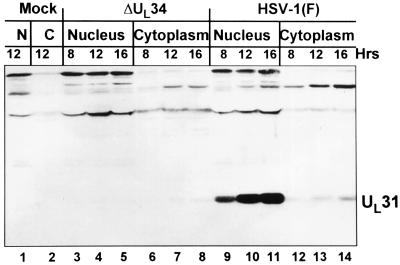
Figure 5.
Immunoblot of electrophoretically separated cell fractionates of rabbit skin cells infected with R5601(ΔUL34) or wild-type HSV-1 (F). Rabbit skin cells (1 × 10⁶) were exposed to R5601 or HSV-1(F) at 5 pfu per cell and incubated at 37°C. Cells were harvested at time points of 8, 12, and 16 h after infection, and cell fractionation was done as described in Materials and Methods. The cytoplasmic and nuclear fractions obtained were resuspended in 1× SDS disruption buffer and subjected to electrophoresis on a denatured polyacrylamide gel (12%). After transfer to a nitrocellulose membrane, the separated infected cell proteins were reacted with the polyclonal antibody to UL31. Lanes 1 and 2, nuclear and cytoplasmic fractions of mock-infected cells. Lanes 3–8, nuclear fractions (lanes 3–5) and cytoplasmic fractions (lanes 6–8) of cells infected with R5601 and harvested at 8 h (lanes 3 and 6), 12 h (lanes 4 and 7), and 16 h (lanes 5 and 8) after infection. Lanes 9–14, nuclear (lanes 9–11) and cytoplasmic (lanes 12–14) fractions of cells infected with HSV-1(F) virus, in the same arrangement as that in lanes 3–8.

