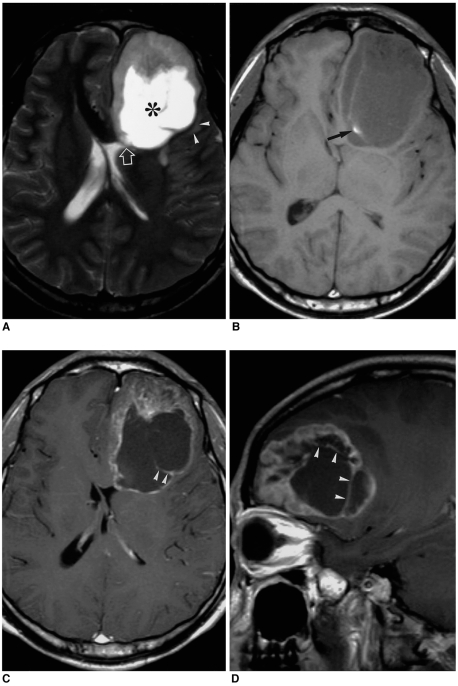
Fig. 1.
A 21-year-old man with supratentorial intraparenchymal ependymoma.
A. Axial T2-weighted spin-echo (SE) image (4500/96) depicts a large tumor with an extensive cystic component (asterisk) in the left frontal lobe. The lesion focally abuts the adjacent frontal horn of the lateral ventricle (open arrow), and there is mild peritumoral edema (arrowheads).
B. Axial T1-weighted SE image (500/12) demonstrates an area of focal hyperintensity within the tumor (arrow),respresenting intratumoral hemorrhage. Due, presumably, to its high protein content, the cystic component appears isointense to gray matter.
C, D. Axial (C) and sagittal (D) contrast-enhanced T1-weighted SE images (500/12) show heterogeneous enhancement and a well-defined tumor margin. The cystic portion is multi-septated and enhanced (arrowheads).

