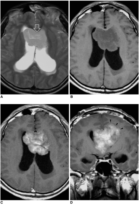
Fig. 2.
A 34-year-old man with supratentorial transependymal ependymoma.
A. Axial T2-weighted SE image (5000/99) depicts a slightly hyperintense, large tumor located within the frontal horns of both lateral ventricles (open arrow), which are dilated.
B. Axial T1-weighted SE image (600/12) shows that the tumor is slightly hypointense and has a lobulated margin.
C, D. Axial (C) and coronal (D) contrast-enhanced T1-weighted SE images (500/12) depict parenchymal invasion adjacent to the left frontal horn (arrowheads), with heterogeneous enhancement.

