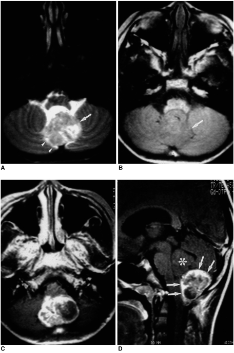
Fig. 3.
A 7-year-old boy with infratentorial ependymoma.
A, B. Axial T2-weighted SE image (A) (3000/100) and axial T1-weighted SE image (B) (650/25) demonstrate, respectively, hyper- and isointensity of the large tumor (arrows) seen at the midline of the posterior fossa. Associated mild peritumoral edema (arrowheads) is present.
C. Axial contrast-enhanced T1-weighted SE image (650/25) reveals heterogeneous enhancement and a cystic component.
D. Sagittal contrast-enhanced T1-weighted SE image (450/25) shows that the tumor extends caudally through the foramen of Magendie. Its intraventricular portion (asterisk) is not enhanced, whereas the caudally extended portion (in the cisterna magna) shows relatively intense enhancement (arrows).

