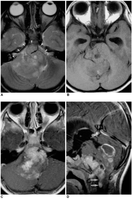
Fig. 4.
A 3-year-old boy with infratentorial ependymoma.
A. Axial T2-weighted SE image (4500/96) depicts a large slightly hyperintense mass in the fourth ventricle. The mass extends to the right cerebellopontine angle and prepontine cistern through both lateral recesses and the foramen of Luschka (arrowheads), encasing a linear signal void (arrow) thought to be a vascular structure.
B. T1-weighted SE image (500/12) reveals the presence of a small area of focal high signal intensity (open arrow) within the mass, suggesting intratumoral hemorrhage.
C, D. Axial (C) and sagittal (D) contrast-enhanced T1-weighted SE images (500/12) show heterogeneous enhancement, with multiple cystic components and associated peripheral rim enhancement. The mass extends to the cerebellopontine angle bilaterally, and to the level of the upper cervical cord caudally (arrows).

