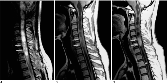
Fig. 5.
A 30-year-old woman with spinal ependymoma of the cervical cord.
A, B. Sagittal T2-weighted SE image (A) (4000/120) shows that the mass at the level of C-5 to C-6 (arrows) is heterogeneously hyperintense, while T1-weighted image (B) (671/12) shows hypointensity. Associated rostral and caudal cysts are also visible.
C. Sagittal contrast-enhanced T1-weighted SE image (671/12) shows homogeneous enhancement and a well-defined, enhanced border (arrows). Extensive associated rostral and caudal cysts (asterisks) extend from the level of the foramen magnum to T-3.

