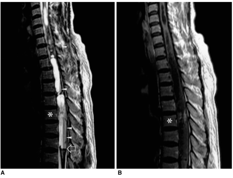
Fig. 6.
A 50-year-old woman with spinal ependymoma at the thoracic level.
A. Sagittal T2-weighted SE image (3500/108) depicts an extensive heterogeneous lesion of mixed signal intensity in almost the entire spinal cord. Rostral and caudal cysts with inner multifocal fluid fluid levels (arrowheads) are extensive, and after previous hemorrhage, hemosiderin has been deposited. In addition, a dark line suggesting hemosiderin deposition (open arrow) is noted along the cord and the bottom of the caudal cyst. Asterisk indicates the T-5 level of the small enhancing tumor seen in B.
B. Sagittal contrast-enhanced T1-weighted SE image (500/14) depicts a small area of slightly heterogeneous enhancement at the T-5 level of the spinal cord (asterisk).

