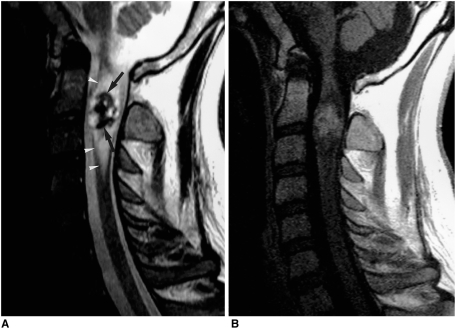
Fig. 7.
A 27-year-old man with ependymoma of the cervical cord.
A. Sagittal T2-weighted SE image (3500/120) shows that in the upper cervical cord, a heterogeneously intense mass with a low signal intensity rim (arrows) at its upper and lower margins is present. Associated peritumoral edema (arrowheads) is also apparent.
B. Sagittal contrast-enhanced T1-weighted SE image (500/15) of the tumor depicts heterogeneous mild enhancement.

