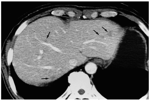
Fig. 1.
Focal eosinophilic necrosis of the liver in a 51-year-old man with gastric cancer and peripheral eosinophilia (11.7%). Contrast-enhanced CT scan obtained during the portal venous phase shows that in both hepatic lobes, several non-spherical lesions (arrows) with a fuzzy margin are present, and there is subtle hypoattenuation without rim enhancement.

