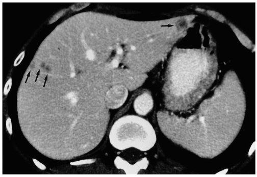
Fig. 3.
Metastases from rectal cancer first evident at follow-up CT performed seven months after surgery in a 62-year-old man. Contrast-enhanced helical CT scan obtained during the portal venous phase shows small hypoattenuating lesions (arrows) with an obvious rim- or target-like enhancement pattern.

