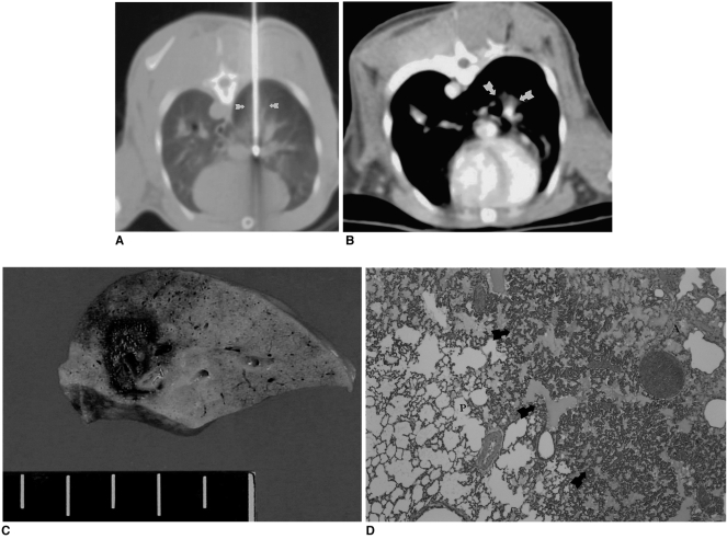
Fig. 1.
Radiofrequency ablation in a rabbit in the radiofrequency group.
A. Computed tomographic section obtained during radiofrequcy ablation. The right lower lobe is penetrated posteriorly by an electrode along which an ovoid opacity extends (arrows).
B. Contrast-enhanced CT scan obtained after the procedure depicts nonenhancing opacity (arrows) in the central portion of the right lobe. Note good enhancement of the pulmonary vessels in the area of ablation.
C. Gross specimen demonstrates an 8-mm central zone of coagulation necrosis surrounded by a dark-brown, 1-mm-thick hemorrhagic rim.
D. Microscopic image of the central ablation zone (A) shows that this contains pyknotic nuclei and eosinophilic cytoplasm, and that hemorrhagic congestion (arrows) has occurred at the border between the ablation area and normal pulmonary parenchyma (P).

