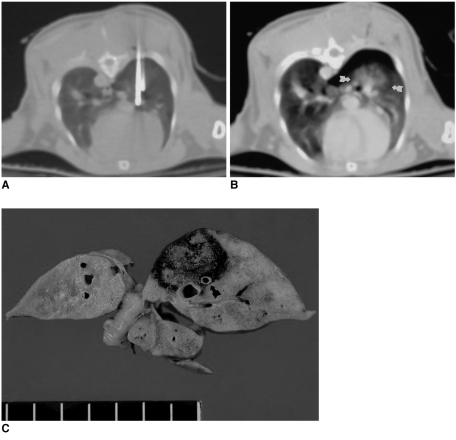
Fig. 2.
Saline-enhanced radiofrequency ablation in a rabbit in the saline-enhanced radiofrequency group.
A. A 17-gauge RF electrode and a 21-gauge coated Chiba needle have been placed in the right lower lobe of the lung. To maximize the effect of saline infusion, the tip of the needle is positioned 3mm posterior to the tip of the electrode.
B. Axial CT scan obtained after RF application shows a rounded opacity 12 mm in diameter (arrows), corresponding to coagulated alveoli and pneumothorax.
C. Gross specimen demonstrates a dark-brown area of coagulative necrosis surrounded by a peripheral hemorrhagic rim. Note the somewhat lobulated margin of the ablation zone, with focal sparing of the pulmonary artery (arrow).

