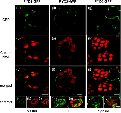
Fig. 5.
Localization of PYD1-GFP, PYD2-GFP and PYD3-GFP in mesophyll cells of stably transformed Arabidopsis. (a) PYD1-GFP, GFP fluorescence signal (green); (b) PYD1-GFP, chlorophyll autofluorescence signal (red); (c) PYD1-GFP, overlay of GFP and chlorophyll fluorescence signals; (d) PYD2-GFP, GFP fluorescence signal; (e) PYD2-GFP, chlorophyll autofluorescence signal; (f) PYD2-GFP, overlay of GFP and chlorophyll fluorescence signals; (g) PYD3-GFP, GFP fluorescence signal; (h) PYD3-GFP, chlorophyll autofluorescence signal; (i) PYD3-GFP, overlay of GFP and chlorophyll fluorescence signals. (j–l) AMK2-GFP, plastid control: GFP fluorescence signal (green), chlorophyll autofluorescence signal (red) and overlay (Lange et al., 2008); (m, n) mgfp4-ER, endoplasmic reticulum (ER) control: GFP fluorescence signal (green) and overlay with chlorophyll autofluorescence signal (red) (Haseloff 1998); (o, p) GFP only, cytosolic control: GFP fluorescence signal (green) and overlay with chlorophyll autofluorescence signal (red); Bar, 8 µm.

