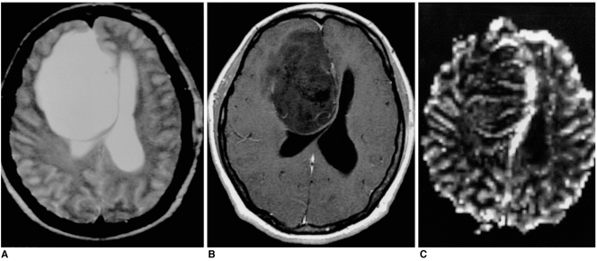
Fig. 2.
Low-grade astrocytoma in a 39-year-old woman. Conventional T2-weighted MR image (A) reveals the presence of a large infiltrative mass with very high signal intensity in the right frontal lobe. Enhanced T1-weighted MR image (B) shows no contrast enhancement. rCBV map (C) demonstrates homogeneous low rCBV, the ratio of which was 1.82.

