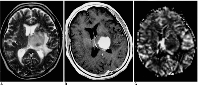
Fig. 3.
Diffuse large B-cell lymphoma in a 68-year-old woman. Conventional T2-weighted MR image (A) depicts a round mass with intermediate signal intensity in the left thalamus. Enhanced T1-weighted MR image (B) demonstrates homogeneous intense enhancement, while rCBV map (C) shows homogeneous low rCBV (ratio, 1.72).

