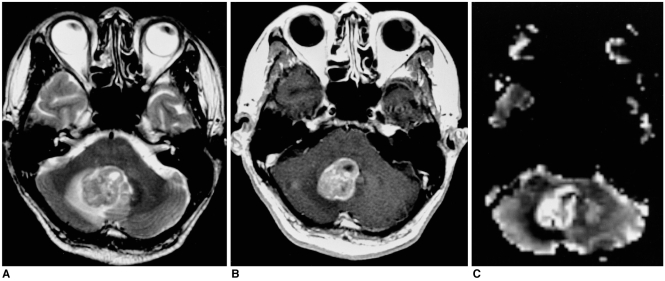
Fig. 4.
Metastatic squamous cell carcinoma of the lung in a 56-year-old man. Conventional T2-weighted MR image (A) demonstrates a lobulated mass with intermediate signal intensity in the right cerebellum. Enhanced T1-weighted MR image (B) shows relatively strong enhancement. rCBV map (C) depicts the tumor's relatively high rCBV, the ratio of which was 7.88.

