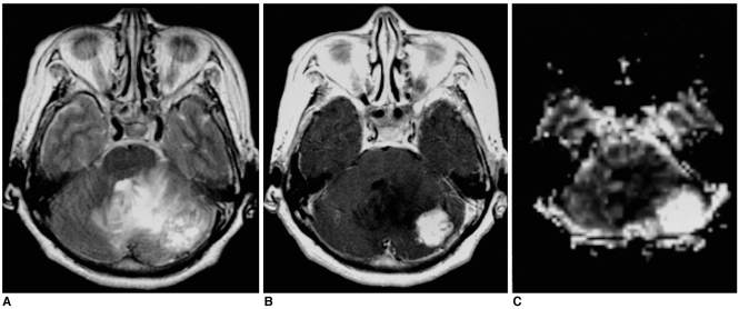
Fig. 5.
Solid hemangioblastoma in a 62-year-old woman. Conventional T2-weighted MR image (A) shows that in the left cerebellum, a lobulated mass with inhomogeneously high signal intensity is present. Enhanced T1-weighted MR image (B) shows strong enhancement. rCBV map (C) demonstrates very high rCBV (ratio, 40.75).

