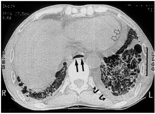
Fig. 10.
Esophageal involvement in a 59-year-old man with scleroderma. Lung window of thin-section (1-mm collimation) CT scan obtained at the level of the liver dome shows a dilated esophagus (arrows), a moderate amount of pericardial effusion (open arrows), and left pleural effusion (curved arrows). Also note the occurrence of pulmonary change consisting of irregular linear opacities, ground-glass opacities, and honeycombing.

