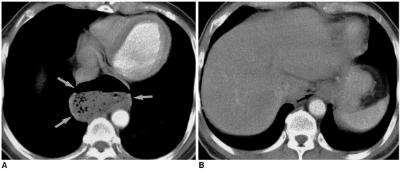
Fig. 17.
Epiphrenic esophageal diverticulum in a 63-year-old man.
A. Mediastinal window of enhanced (7-mm collimation) CT scan obtained at the level of the suprahepatic inferior vena cava shows that outpouching (arrows) from the esophagus contains an air-fluid level.
B. CT scan obtained at the level of the liver dome shows that the position of the esophagogastric junction (arrow) is normal.

