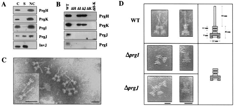
Figure 3.
prgI and prgJ are required for needle structure formation. (A) Western-blot analysis of whole cell lysate (C), culture supernatant (S), and postCsCl needle complex fractions (NC) isolated from WT Salmonella (TK328). The amount of C and S fractions loaded represents equivalent amounts of total culture volume (0.2 ml), the NC represents 25 ml of culture. The blot was probed with polyclonal antiserum against PrgH, PrgI, PrgJ, PrgK, and InvJ as indicated. (B) Western blot analysis of PreCsCl NC fractions isolated from WT and prg deletion strains: WT (TK328), ΔH (TK334), ΔI (TK335), ΔJ (TK336), ΔK (TK337), ΔH-K (TK338). The blot was probed with polyclonal antiserum against PrgH, PrgI, PrgJ, and PrgK as indicated. (C) Electron micrographs of NC isolated from WT (TK328). Complexes came from same fraction (NC) analyzed in A. Samples were negatively stained with 2% PTA (pH 7.2) and observed under a JEM-1200EXII transmission electron microscope. Approximate sizes of the NC are listed in the schematic (D) (n = 30). (Scale bar, 50 nm.) (D) Electron micrographs of partial NC isolated from WT (TK328), ΔprgI (TK335), and ΔprgJ (TK336) deletion mutants. A schematic comparing the size and morphology of these structures to WT structures is also shown. Approximate sizes are given in schematic (n = 30). (Scale bars, 50 nm and 25 nm.)

