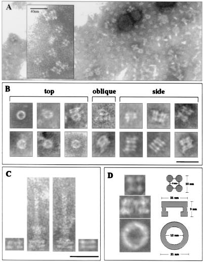
Figure 5.
PrgH and PrgK form the inner membrane rings of the NC. (A and B) Representative electron micrograph images of PrgH/PrgK complexes from recombinant E. coli (DH5α) expressing prgH/prgK (pTK42) or prgHIJK (pTK53). (B) Electron micrographs of individual ring structures (top), rectangular structures (side), and oblique view of same structures. (Scale bar, 50 nm.) (C) Comparison of two different side views of the PrgH/K complex with NC isolated from WT Salmonella. Note that the differences seen in these structures are reflected in the differences seen in the base structure of the NC. Scale bar, 50 nm. (D) Schematic comparing the approximate size and shape of the isolated Prg components: tetramer, 10 nm (n = 25); rectangular (side), width 21 nm, height 9 nm (n = 27); ring (top), outer diameter 21 nm, inner diameter 12 nm (n = 30).

