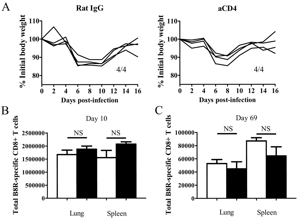
Figure 1. CD4 help does not affect the initial disease and CD8+ T cell kinetics during VV infection.
(A) Weight loss after infection was measured. Numbers indicate surviving mice. (B,C) The VV B8R-specific CD8+ T cell response was measured by tetramer staining on day 10 (B) and day 69 (C) p.i. Total numbers of CD8+ tetramer+ cells in each organ are graphed. White bars: un-depleted, black bars: CD4-depleted. Representative data from two independent experiments with 4 mice per group are shown. Error bars indicate SEM. NS: not significant.

