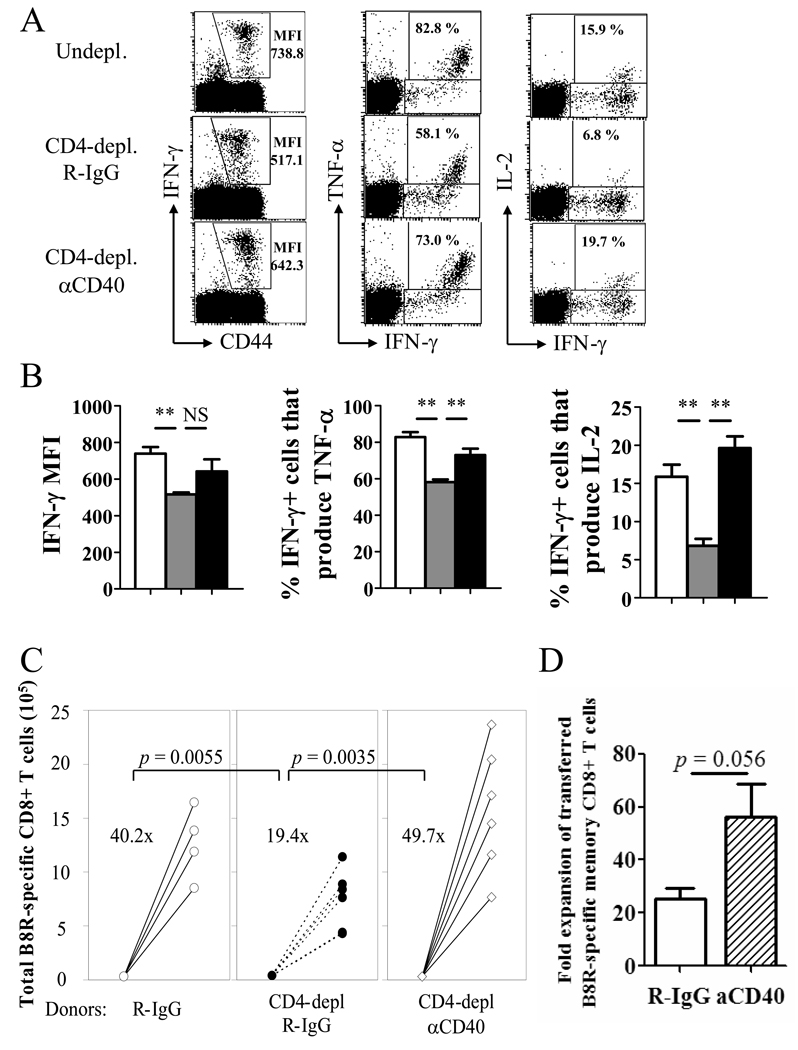
Figure 5. Providing CD40 stimulation during priming boosts memory cell function in the absence of CD4 help.
Mice were infected with VV-WR and were un-depleted, CD4-depleted/R-IgG-treated, or CD4-depleted/anti-CD40-treated (d1, 7). A, Cytokine production by VV-specific memory CD8+ T cells was analyzed by ICCS. Plots are gated on CD8+ cells. Numbers indicate IFN-γ MFI of CD8+ T cells producing IFN-γ (left), or percentage of IFN-γ+ cells that produce TNF-α (middle) or IL-2 (right). B, Graphed representation of (A). White bars: un-depleted, grey bars: CD4-depleted/R-IgG-treated, black bars: CD4-depleted/anti-CD40-treated. Representative data from two independent experiments with 3–4 mice per group are shown. Error bars indicate SEM. NS: not significant. * p > 0.05, ** p > 0.01. C, The recall response by VV-specific memory CD8+ T cells was analyzed by adoptive transfer and challenge. Treatments of donor mice are indicated below. Numbers indicate average fold expansion of VV-specific memory cells for each group. Each circle represents an individual recipient. D, Recall response of helped (undepleted) memory cells that received R-IgG or anti-CD40 during priming is graphed. Fold expansion of transferred (CD45.2+) memory cells is shown (error bars indicate SEM). Representative plots are shown from two independent experiments; consisting of 3–4 mice per group are shown. In C and D, 4 donor mice per group, and 4–6 recipients per group were used.

