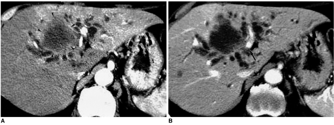
Fig. 10.
Arterioportal shunt secondary to cholangiocarcinoma.
A. CT scan obtained during hepatic arterial phase shows diffuse hyperattenuation at the left lobe of the liver, as well as a large hypoattenuating mass (arrowheads) and dilated bile ducts.
B. Portal venous phase image demonstrates that parenchymal enhancement has returned to nearly normal. Note that the left portal vein is encased by the tumor (arrow).

