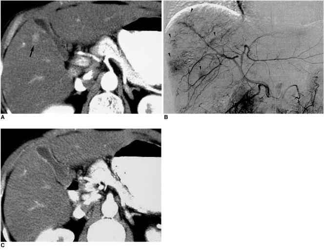
Fig. 11.
Nodular-shaped nontumorous arterioportal shunt mimicking hepatic tumor in cirrhotic liver.
A. CT scan obtained during hepatic arterial phase shows a nodular hyperattenuating lesion (arrow) in the right lobe of the liver. Note the subtle, tiny portal branch inside the lesion.
B. Subsequent hepatic angiogram shows multiple patchy areas of staining in the liver (arrowheads), with early opacification of the portal veins.
C. Follow-up CT scan obtained during hepatic arterial phase shows no hyperattenuating lesion in the same area, indicating that the lesion depicted in (A) was a nontumorous arterioportal shunt.

